Sonographie (Ultraschall) in Berlin Treptow-Köpenick
Dr. med. Karolin Stein

Was ist die Sonographie (Ultraschall)?
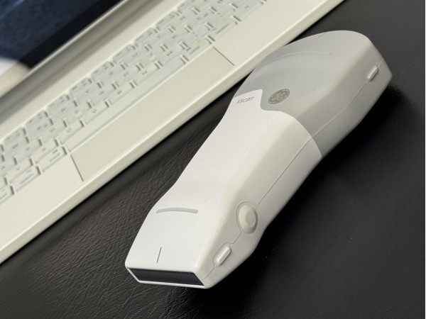
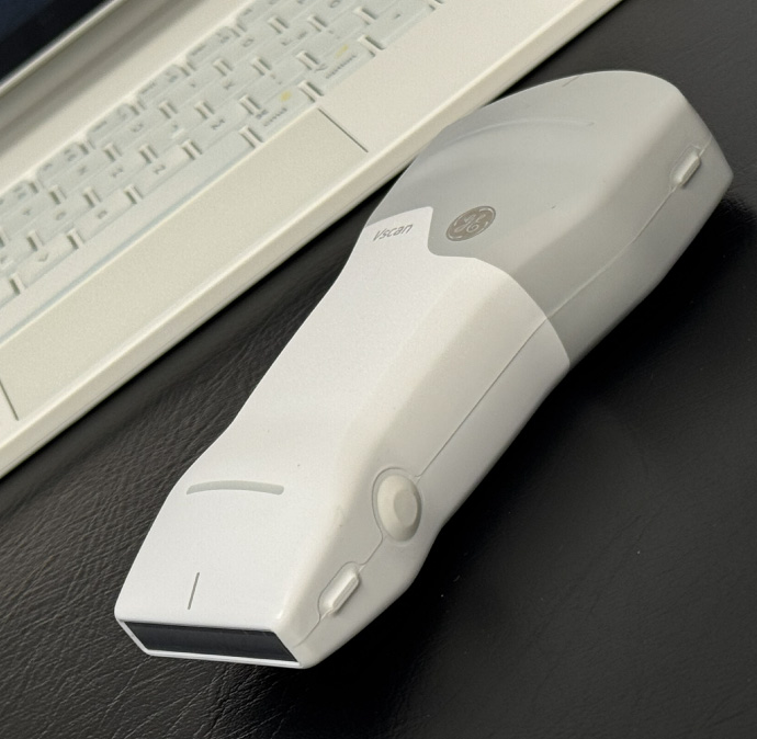
Sonographie in der Orthopädie – häufig auch Muskuloskelettaler Ultraschall genannt – ist ein modernes, strahlenfreies Bildgebungsverfahren, das zur Diagnose und Verlaufskontrolle von Erkrankungen des Bewegungsapparates eingesetzt wird. Mit Hilfe hochauflösender Ultraschallgeräte können Muskeln, Sehnen, Bänder, Gelenke und Weichteile in Echtzeit dargestellt werden.
Typische Einsatzbereiche
- Sehnen- und Muskelverletzungen: z. B. Achillessehnenentzündung, Muskelrisse, Sehnenscheidenentzündungen
- Gelenknahe Beschwerden: Darstellung von Schleimbeuteln, Gelenkergüssen, Bandverletzungen
- Schulterdiagnostik: z. B. Impingement-Syndrom, Rotatorenmanschettenverletzungen
- Hüft- und Kniebeschwerden: Erkennung von Entzündungen, Ergüssen, Strukturveränderungen
- Beurteilung von Weichteilschwellungen: Zysten, Lipome, Hämatome
- Gezielte, ultraschallgesteuerte Infiltrationen: z. B. an Schulter, Knie, kleinen Gelenken
-
Strahlenfrei und schonend
Die Untersuchung erfolgt ohne Röntgenstrahlung und ist daher besonders schonend – ideal auch für Kinder und Schwangere.
-
Dynamische Untersuchung
Bewegungsabläufe können in Echtzeit dargestellt und funktionelle Zusammenhänge direkt beurteilt werden.
-
Hohe Detailgenauigkeit
Oberflächennahe Strukturen wie Sehnen, Muskeln, Bänder und Gelenke lassen sich präzise und differenziert darstellen.
-
Sofortige Befundung
Die Beurteilung der Befunde erfolgt direkt während der Untersuchung, ohne Wartezeit.
-
Optimale Unterstützung bei Injektionen
Injektionen können unter Sicht exakt platziert werden, da Nadeln sonographisch präzise geführt werden.
Häufige Fragen zur Sonographie (Ultraschall)
Ist die Ultraschalluntersuchung schmerzhaft?
Nein. Die Sonographie ist eine schmerzfreie Untersuchungsmethode. Der Schallkopf wird mit leichtem Druck über die Haut geführt. In der Regel empfinden Patientinnen und Patienten die Untersuchung als unkompliziert und gut verträglich.
Muss ich mich auf die Ultraschalluntersuchung vorbereiten?
In der Regel ist keine besondere Vorbereitung notwendig. Die Untersuchung kann direkt im Rahmen des Termins durchgeführt werden. Gegebenenfalls bitte ich Sie, die zu untersuchende Körperregion frei zugänglich zu machen.
Wie lange dauert eine Ultraschalluntersuchung?
Die Dauer hängt von der Fragestellung und der zu untersuchenden Region ab. In der Regel dauert eine orthopädische Ultraschalluntersuchung etwa 10 bis 15 Minuten inklusive Befundbesprechung.
Kann Ultraschall ein MRT ersetzen?
In vielen Fällen liefert die Sonographie bereits aussagekräftige Befunde, insbesondere bei Beschwerden der Weichteile wie Muskeln oder Sehnen. In bestimmten Situationen kann jedoch eine weiterführende Bildgebung (z. B. MRT) notwendig sein. Dies kläre ich individuell mit Ihnen.
Kann Ultraschall auch zur Verlaufskontrolle eingesetzt werden?
Ja. Da Ultraschall ohne Strahlenbelastung auskommt, eignet sich die Untersuchung sehr gut zur Verlaufskontrolle, beispielsweise zur Beurteilung des Heilungsverlaufs oder des Therapieerfolges.
Erhalte ich die Ergebnisse direkt?
Ja. Die Befunde liegen unmittelbar nach der Untersuchung vor und werden im Anschluss verständlich mit Ihnen besprochen. Daraus ergeben sich direkt die nächsten diagnostischen oder therapeutischen Schritte.